
As a key member of the National Imaging Facility (NIF), our specialist facilities and expertise at Swinburne Neuroimaging is open to all Australian researchers and enables them to tackle today’s most pressing brain-related research questions.
We support research that probes the rapid dynamics of human brain function and underpins behaviour, cognitive development and mental health across the lifespan.
We're the sole single-site human imaging facility in Australia and New Zealand to offer magnetoencephalography (MEG) and magnetic resonance imaging (MRI).
In addition, we have a wide range of additional and complementary capabilities.


MEG, MRI and EEG equipment
The Neuroimaging facility here at Swinburne is the only single-site human imaging facility in Australia and New Zealand to offer magnetoencephalography (MEG), magnetic resonance imaging (MRI) and electroencephalography (EEG).
MEG allows us to measure the brain at the speed at which neural activity occurs — that's in the order of milliseconds. By contrast, it takes seconds for blood to flow in the brain. This slower speed limits even the fastest MRI scanners. MEG is a safe and non-invasive brain imaging technique that offers high temporal precision that’s able to measure very small magnetic fields that the brain’s electrical activity produces.
This specialised equipment can also detect and accurately localise rapid changes in brain activity and connectivity. We house an Elekta/Neuromag 306 channel MEG system in a highly magnetically shielded room to yield extremely clean, low noise data.
MEG helps aid research in many areas, such as traumatic brain injury, dementia, stroke and autism. We’re one of only two sites in the Southern Hemisphere with the magnetoencephalography (MEG) equipment.
In addition our electroencephalography machine is Neuroscan high density MEG and MRI compatible.
Our neuroimaging research is already creating a real impact on people's lives — and we've barely started.
MEG’s temporal precision is important as it lets us investigate single, rapid processes. This is crucial when we’re looking at the timing of thousands of coordinated processes.
These steps must occur in exactly the right sequence for normal brain function.
Compare this with how a car engine’s fuel injection system works.
A car engine needs to fire each cylinder rapidly. The fuel injection system does this at a precise moment, relative to the other cylinders. When this timing degrades, severe damage can result.
The human brain is like an engine with hundreds of thousands of cylinders, all in harmony with one another.
Our MEG scanner provides a unique means to examine changes in neural activity and timing.
These could underpin conditions such as:
- epilepsy
- traumatic brain injury
- dementia
- stroke
- anxiety
- depression.
In addition, MEG promises to give new insights into developmental conditions like:
- autism
- attention deficit hyperactivity disorder (ADHD).
Magnetic resonance imaging (MRI) is a medical imaging technique that uses a powerful magnetic field to obtain very detailed cross-sectional images inside the body and brain.
Our Siemens 3 Tesla whole-body MRI scanner offers extremely fine-grained structural and functional images of the entire body. It also has specialised equipment for human brain imaging.
In May 2020, we upgraded our Siemens Tim Trio 3T MRI to a 3T Prisma Fit.
Our MRI system has the latest scanning sequences installed. This gives researchers access to world-leading techniques to image the human brain’s structure, function and connectivity.
Electroencephalography (EEG) records the brain's electrical activity, with electrodes placed on the scalp.
Our Neuroimaging facility currently has:
- 3 laboratories with 64 channel Neuroscan Synamps2/RT systems (2 of them with Steady State Topography)
- 1 laboratory with a 40 channel Neuroscan NuAmp System
- a magnetic resonance imaging (MRI) compatible 132 channel Neuroscan system
- 3 magnetoencephalography (MEG) compatible 64 channel ANT systems
- a 'Babylab' (128 channel EGI NetAmp system with infant HydroCel Geodesic Sensor nets).
Used alone, EEG provides accessible, high quality recording of brain activity. EEG can also be combined with MRI or MEG to maximise sensitivity and localisation of brain activity.
-
 upgrade to a Siemens 3T Prisma (completed May 2020)") Swinburne’s magnetic resonance imaging (MRI) upgrade to a Siemens 3T Prisma (completed May 2020)
Swinburne’s magnetic resonance imaging (MRI) upgrade to a Siemens 3T Prisma (completed May 2020) -
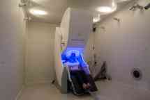
 A woman having a Magnetoencephalography scan
 -
 scanner") Swinburne’s Elekta Neuromag TRIUX magnetoencephalography (MEG) scanner
Swinburne’s Elekta Neuromag TRIUX magnetoencephalography (MEG) scanner
 upgrade to a Siemens 3T Prisma (completed May 2020)")

 scanner")
Other equipment at the Swinburne Neuroimaging facility
Our state-of-the-art neuroimaging facility boasts the most modern technology for measuring brain activity.
In addition to our MEG and MRI equipment, we have a wide range of additional and complementary capabilities. Among them are:
- transcranial magnetic stimulation or TMS (Magstim 200 with BiStim Controller, Magstim single pulse)
- eye tracking (Eyelink 1000 systems integrated with MRI and MEG)
- peripheral psychophysiology
- integrated experimental presentation hardware and software
- powerful cloud-based data management and analysis platforms.
Our imaging systems are all configured with:
- experimental and stimulus presentation computers
- high quality visual and auditory stimulus delivery
- behavioural response devices (button boxes and joysticks)
- computers for data acquisition connected to networked storage.
High-speed eye tracking (Eyelink 1000) is available for MRI and MEG experiments.
Swinburne Neuroimaging is also equipped with technology for non-invasive brain stimulation. This includes transcranial magnetic stimulation (TMS) and transcranial direct current stimulation (tDCS).
Our experience helps us to develop solutions. Where needed, we can use other technologies within the imaging labs.
Swinburne Neuroimaging has recently transitioned its computing to the OzStar Supercomputer. In this way, we offer the highest performance analytic pipelines.
Our user-friendly, locally hosted virtual computing environments enhance the supercomputing environment. The systems are suitable for interactive analysis, training and tutorials.
We offer a wide range of neuroimaging analysis software and processing pipelines. We store data in Brain Imaging Data Structure (BIDS) format.
We're connected to national computing infrastructure, such as Nectar. As a result, we can provide support for national collaborative research.
-
Swinburnes Neuroimaging projects and case studies
Our highly experienced team here at Swinburne Neuroimaging (SNI) facility develop cutting-edge brain imaging that supports our pioneering research. Explore a selection of our recent projects and read our case studies.

Our highlights

Towards Curing Epilepsy by Tracking the Eye of the Storm
Dr Miao Cao shares how a breakthrough in 3D Field Imaging offers new hope to those living with uncontrolled, life-threatening seizures.
-
- Health
World-first disease progression model offers new hope for muscular dystrophy patients
Patients with a common type of muscular dystrophy will benefit from a world-first AI powered disease progression model with ‘life-changing’ implications, using advanced MRI imaging at Swinburne University of Technology.
Friday 05 September 2025 -
- Technology
- Health
- Science
The new non-invasive brain scan techniques giving seizure sufferers a new lease on life
Swinburne University of Technology’s cutting-edge non-invasive investigations for epilepsy are giving seizure sufferers like Stephenie Evans a second chance at a happy and healthy life.
Read more (The new non-invasive brain scan techniques giving seizure sufferers a new lease on life )Monday 16 October 2023


Supporting your research
We can help you with all steps of your research such as, project conception, project design and logistical planning, advice on research ethics, advice on data management and providing expert training in data analysis. Contact the team to discuss your project via neuroimaging@swinburne.edu.au.
Once contact has been made, you can begin the application.
SNI Access and Support Scheme
The Swinburne Neuroimaging (SNI) Access and Support Scheme is designed to promote, facilitate and support world-class innovation and research in neuroimaging. Applications for the following grants are now being accepted:
- SNI Early Career Research Grant
- SNI Feasibility Grant
- SNI Enhancing Capability Grant
- SNI Top-Up Grants for Innovative Research
Our people
-
Associate Professor David White
Director, Swinburne Neuroimaging -
Dr Rachel Oliver
Operations Manager, Swinburne Neuroimaging -
Dr Will Woods
MEG Fellow -
Dr Miao Cao
Informatics Fellow -
Dr Matthew Hughes
MRI Fellow -
Professor Chris Plummer
Adjunct Professor -
Mr Simon Vogrin
Adjunct Researcher -
Professor Alan Pearce
Adjunct Professor
Explore our news
Contact the Swinburne Neuroimaging team
Whether you’re a PhD student, media, or an organisation looking to access our facility or partner with us, please contact Dr Rachel Oliver (Batty), Operations Manager on +61 3 9214 8006 or via neuroimaging@swinburne.edu.au.