The inside of cells as never seen before

In Summary
- This article featured in Swinburne’s 2017 ‘Research Impact’ magazine, produced in association with Nature Publishing Group.
Professor Sarah Russell, an immunologist at Swinburne’s Centre for Micro-Photonics, leads the Cell Biology lab. Her group fine-tuned a super-resolution microscopy method known as STORM, which has allowed researchers to capture clear images to the tens of nanometres.
STORM is dependent on antibodies to ‘hold’ colour molecules, so that the parts of the cell researchers wanted to see under the microscope can be tagged. But using antibodies brought technical problems that interfered with accuracy and created blurring of the images.
“You’re restricted by chemistry. The antibody is a whacking big molecule, and when you are homing in on these very precise locations, the last thing you need during imaging is a great big thing wobbling around, increasing the risk of error,” says Professor Russell.
The turning point was when Ye Chen, a Post-Doctoral Fellow in Professor Russell’s lab developed some tricks with which to substitute the antibody with a smaller molecule, such as a cell membrane dye. Replacing the usual antibody middleman with smaller molecules led to a method that didn’t damage the cell and consistently generated a brighter and stronger signal for the microscope.
The results were images that resembled a finely woven net, showing in crisp detail fibres that make up the structural scaffolding in the cell membrane, in far greater resolution than the original STORM method.
Professor Russell’s group discovered the technique also had the potential to show precisely how fibres are arranged. This clearer picture could provide information on the nuances of fibre organisation in different cells and compartments, providing clues on development and function.
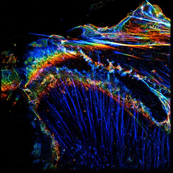
The structural fibres of a cell showing the level of precise detail made possible by the new imaging method.
Being a biologist embedded in the company of physicists in a 16-year-long collaboration between Peter MacCallum Cancer Centre and Swinburne researchers meant that Russell had access to expertise that allowed her to fast-track ideas.
“There is so much physics and chemistry that can be used to improve imaging. This work is a clear indication why it is so valuable to have physicists who already know how to talk to biologists and vice versa,” says Russell.
“At Swinburne we can work together and it’s a fantastic illustration why this collaboration is such a good set-up.”
The results were published in mid-2016 but Russell’s group is already closing in on another imaging technique that makes use of the polarisation of light. Together with the fine-tuned STORM technique, she plans to see both methods applied to monitoring and recording how immune cells respond during an infection in real-time.
“The major effort is to keep tweaking until we get to the stage where these technologies are genuinely valuable for learning about biology,” says Russell. “At the moment there have not yet been many new biological discoveries made based on super-resolution imaging. But nobody is giving up on it and refining the methods will be critical for biological breakthroughs.”