Collaboration critical to breakthrough 3D bioprinting

In Summary
- Swinburne partners in establishing revolutionary bioengineering facility at St Vincent’s Hospital
- PhD candidate develops method to feed cells printed into patients in real time
- Swinburne researchers encouraged to use facility
Swinburne research into how stem cells are ‘fed’ once inside the body could bring surgeons a step closer to regenerating damaged or missing tissue through biological 3D printing techniques.
PhD candidate Lilith Caballero Aguilar is developing methods to control the rate of release of ‘growth factors’ which are necessary for the development of stem cells once they have been implanted into the body.
Controlling this delivery is critical, as stem cells can take six weeks or more to grow into tissue such as cartilage, requiring the growth factors to be slowly released over that time.
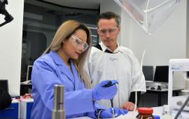
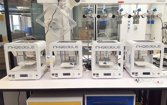
Ms Caballero Aguilar is collaborating with researchers and surgeons at BioFab3D@ACMD, the first bioengineering facility in Australia to be based in a hospital.
Established through a partnership between St Vincent’s Hospital, Swinburne, RMIT, University of Melbourne and University of Wollongong, the facility brings together surgeons, biomedical engineers and biology experts to pioneer innovations such as re-engineered limbs, tissues and nerves.

Ms Caballero Aguilar’s research forms part of two major projects run out of the facility—one focusing on the regeneration of damaged cartilage and the other on the repair of damaged muscle fibres.
Both projects are using advanced technologies to implant materials into the body, including the handheld 3D printer ‘Biopen’ developed by St Vincent’s Hospital and University of Wollongong that permits a surgeon to ‘draw’ biomaterials directly into a patient.
Ms Caballero Aguilar’s research focuses on manipulating polymer materials into suitable release mechanisms for growth factors, which then form part of the 'ink' that is drawn into the body.
She uses the emulsion method of shaking an oil and water solution at an intense rate to create ‘microspheres’. These microspheres are then crosslinked to form a substance that can hold the growth factors.
Collaboration critical
Her supervisor, Swinburne Professor of Biomedical Electromaterials Science Simon Moulton, says the opportunity to collaborate directly with orthopaedic surgeons and muscle specialists at St Vincent’s Hospital has driven the success of the project.
“Without this space, Lilith’s project would be a much smaller project without the translation benefit,” says Professor Moulton.
“It still would be great research done at a very high level, she would have publications and be able to graduate, but working in this collaborative environment, she can achieve all of that, while also having her research go into a clinical outcome that actually has benefit to patients.”
Ms Caballero Aguilar agrees that working alongside not only surgeons, but also other university researchers, has impacted significantly on her research.
“We complement each other. If I have doubt, we can discuss it and reshape the project as we go, which helps to reach a better outcome,” she says.
“At the end of the day, everyone is doing a bit of work in a big project. It feels very rewarding.”
Swinburne researchers interested in viewing or using the facilities at BioFab3D can contact Professor Moulton on smoulton@swin.edu.au.

