
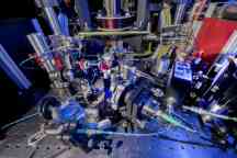
The Optical Sciences Centre is dedicated to pursuing excellence and impact through world-leading research, focusing on both fundamental and applied areas of optical sciences and related areas. Led by Professor David Moss (Director) and Professor Chris Vale and Professor Saulius Juodkazis (Deputy Directors), our centre incorporates staff from the previous Centre for Micro-Photonics and Centre for Quantum and Optical Science to create one of the largest optical research facilities in Australia.
Our vision is to conduct research and development across the full spectrum of research engagement and translation. From fundamental studies in the way light interacts with matter, through to the development of technologies for market, we foster cross-disciplinary collaborations that address scientific challenges in fields encompassing classical and quantum photonics, light-matter interactions, nanotechnology, biomedical and biosciences, quantum gases and quantum materials.
-
-
-

Our study options
We offer opportunities for students wishing to undertake a PhD, masters, honours or undergraduate degree.


-

- Technology
Australian researchers record world’s fastest internet speed from a single optical chip
Researchers have recorded the world’s fastest internet speed from a single optical chip of 44.2 Terabits per second.Friday 22 May 2020

Our publications
The Optical Sciences Centre is a new state-of-the-art research facility with a range of researcher experience, including from the Centre for Micro-Photonics and the Centre for Quantum and Optical Science.
Read publications from the Centre for Micro-Photonics researchers
Read publications from the Centre for Quantum and Optical Science researchers
Centre news
-

- Science
If quantum computing is answering unknowable questions, how do we know they’re right?
A new Swinburne study is tackling the paradox - if quantum computing is answering unknowable questions, how do we know they’re right?
Tuesday 16 September 2025 -

- Technology
- Science
Five surprising ways quantum is changing the world in areas you wouldn’t expect
As the world celebrates the Year of Quantum, here’s five unexpected ways quantum will change the way we operate, explained by Swinburne experts.
Thursday 07 August 2025 -
- Technology
Microsoft, IBM and Google are racing to develop the first useful quantum computer. Ultracold neutral atoms could be the key.
Swinburne University of Technology has been exploring ultracold neutral atoms for two decades.
Monday 30 June 2025 -
- Technology
- Science
Swinburne-led research team demonstrates world’s fastest optical neuromorphic processor
An international team of researchers has demonstrated the world’s fastest and most powerful optical neuromorphic processor for artificial intelligence, which is capable of processing ultra-large scale data
Thursday 07 January 2021


Find more news articles by visiting the Centre for Quantum and Optical Science newsroom and the Centre for Micro-Photonics newsroom.
Explore other research centres
-
Centre for Astrophysics and Supercomputing
-
Centre for Design Innovation
-
Centre for Forensic Behavioural Science
-
Centre for Mental Health and Brain Sciences
-
Centre for Quantum Science and Technology Theory
-
Centre for Social Impact Swinburne
-
Centre for Sustainable Infrastructure and Digital Construction
-
Centre for Transformative Media Technologies
-
Optical Sciences Centre
How you can reach us
Location
Optical Sciences Centre
Swinburne University of Technology
John Street
Hawthorn VIC 3122
Australia
Postal address
Optical Sciences Centre
Swinburne University of Technology
Mail H74
PO Box 218
Hawthorn VIC 3122
Australia
Contact the Optical Sciences Centre
There are many ways to engage with us. If your organisation is dealing with a complex problem, get in touch to discuss how we can work together to provide solutions. Call us on +61 3 9214 8096 or email osc@swinburne.edu.au.